Radiology Tutor
Colon and rectal cancer TNM staging (UICC/AJCC 7th edition)
If there is no evidence of tumour then T0 is used and if the tumour cannot be assessed, TX is used.
Background
Due to the many possible subdivisions and multiple nodal stations, colorectal cancer staging can be daunting. However, whilst important differences in the imaging, anatomy and in the subdivisions (optional) are present, common features mean that the TNM staging of cancers of the colon and the TNM staging of cancers of the rectum are grouped together.
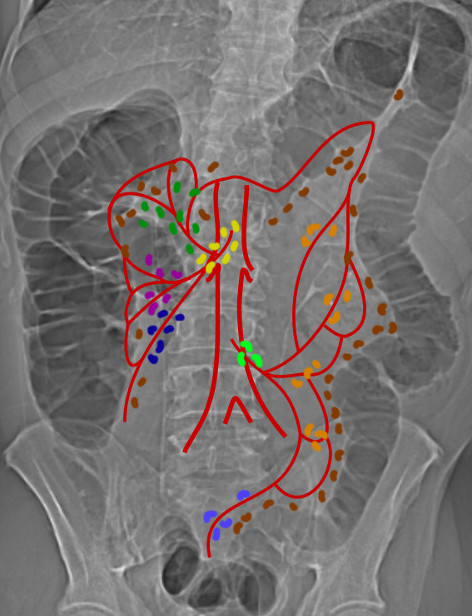
Since T3 tumours extend beyond the muscularis propria and T4 tumours extend beyond the serosa, the anatomical presence or absence of the serosa is important. T3 tumours may extend more than 15 mm beyond the muscularis propria, but if serosa is present close to the muscularis propria, the direct extent as a T3 tumour may be limited, as once the serosa is perforated, the tumour is classified as T4. Parts of the colon that are intraperitoneal are covered with serosa (visceral peritoneum), these are caecum, transverse colon and sigmoid colon and are easily remembered as these are suspended by a mesentery. Visceral peritoneum also covers the ascending and descending colons, but only over their anterolateral surfaces. Posteriorly these structures (beyond their muscularis propria) are covered by adventitia continuous with the retroperitoneum. The rectum is only covered in peritoneum in its midpart anteriorly and upper part anterolaterally. The important boundary in the rectum is the mesorectal fascia as this indicates the circumferential resection margin (CRM, see below).
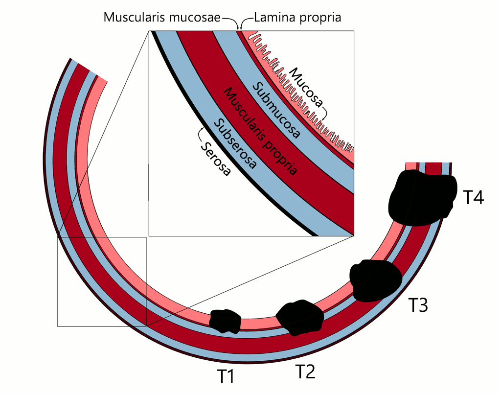
The circumferential resection margin (CRM) corresponds to the site of mesenteric resection (for intraperitoneal structures), the mesorectal fascia (for rectum), or the retroperitoneal dissection margin (ascending and descending colon). For example if a tumour of the transverse colon is resected, but the cut margin of the mesentery contains tumour, this is CRM positive. For the radiologist this is important because in rectal tumours MRI can identify the likely status of the CRM as total mesorectal excision (TME), the surgery of choice, removes all components within the mesorectal fascia and the fascia itself is taken to be the CRM. TNM staging from AJCC staging manual(1) and UICC TNM Classification of Malignant Tumours 7th edition(2):
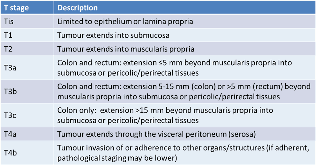
Note that there is no longer a T3d classification for colonic tumours (the 1 mm cut-off has been removed) and now the only T3 subdivisions for rectal tumours are T3a and T3b (only the 5 mm cut-off remains).(3) Also, note that the AJCC has not adopted the T3 subdivision.
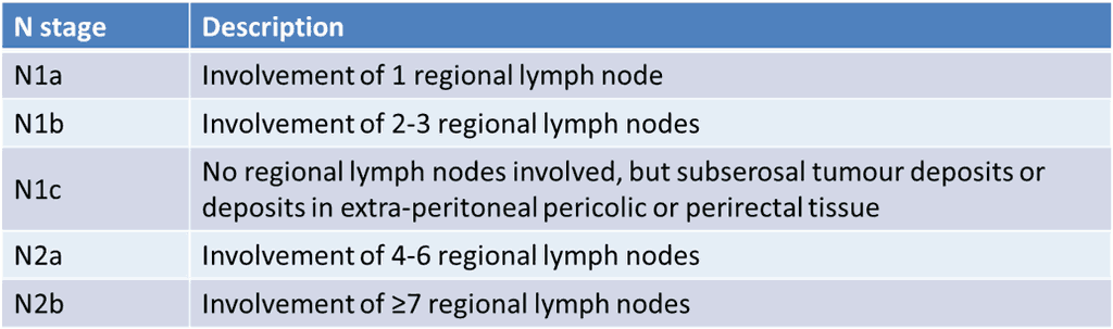
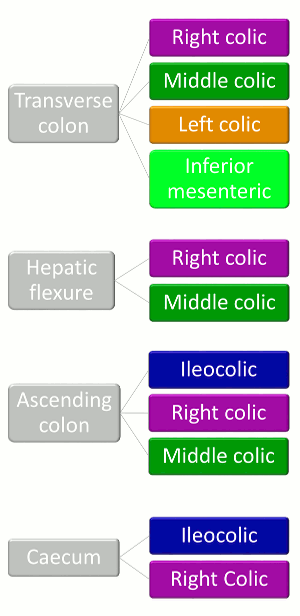
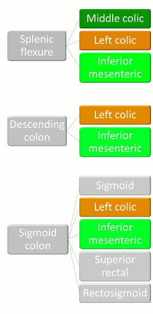
Regional nodes as defined by the AJCC(1):
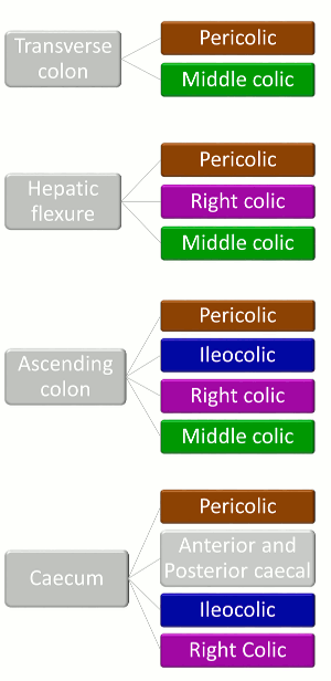
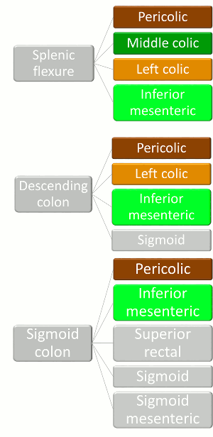
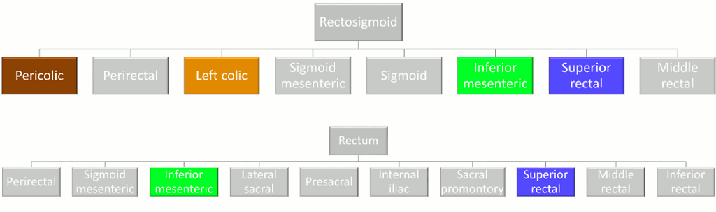
Regional nodes as defined by the UICC(2):

Overall stage for each T and N stage:
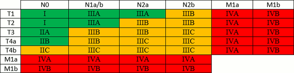
Contrary to the tables found elsewhere in Radiology Tutor, the colour codes here represent prognosis.
References
- S. B. Edge, D. R. Byrd, C. C. Compton, A. G. Fritz, F. L. Greene, and A. Trotti, Eds., AJCC Cancer Staging Manual, 7th ed. 2010. Springer, 2011.
- L. H. Sobin, M. K. Gospodarowicz, and C. Wittekind, Eds., TNM Classification of Malignant Tumours, 7th Edition. Wiley-Blackwell, 2009.
- C. Wittekind, TNM Supplement: A Commentary on Uniform Use, 4th Edition. Wiley-Blackwell, 2012.